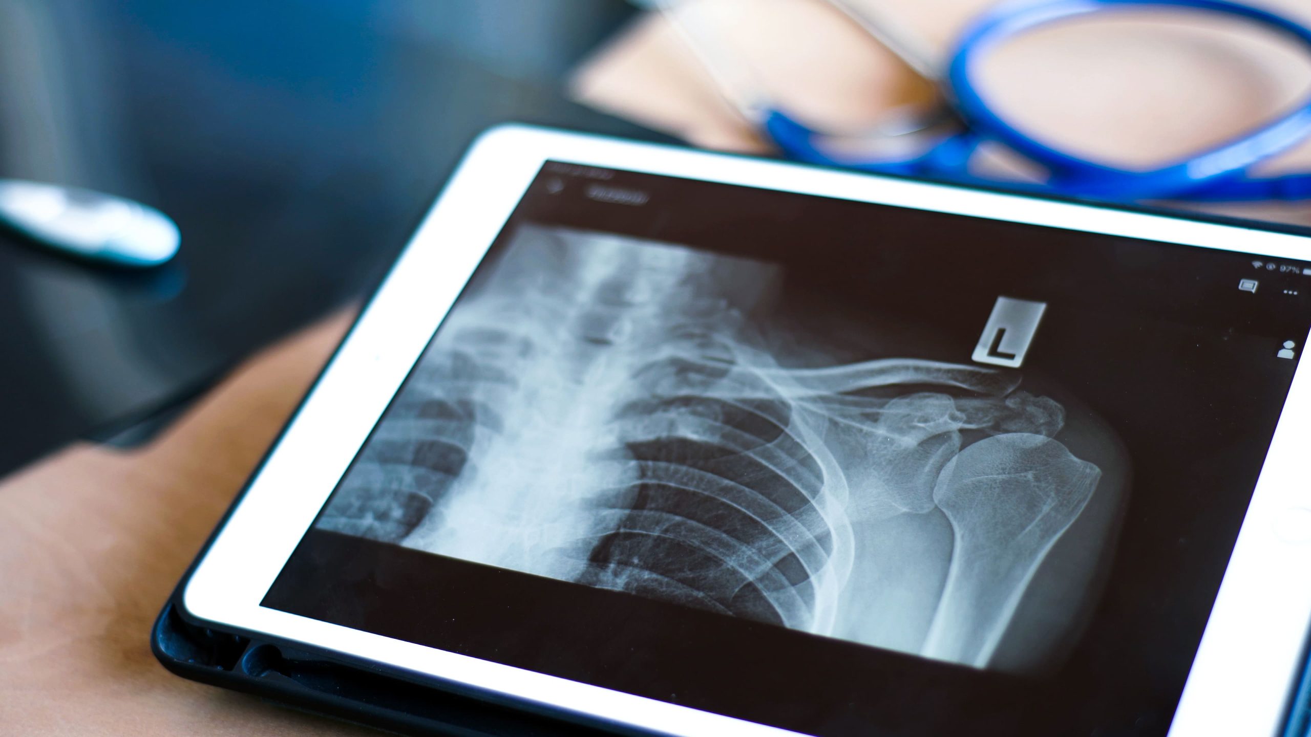

Reaching behind your back to fasten clothing or tuck in a shirt can become difficult when the shoulder joint’s capsule thickens and contracts, a condition known as frozen shoulder. Unlike rotator cuff tears, which may preserve full passive movement while causing weakness and pain with specific activities, frozen shoulder typically restricts motion in every direction. An orthopaedic evaluation uses these dynamic patterns, along with specialised imaging, to guide an accurate clinical diagnosis and construct an appropriate management pathway.
Understanding Frozen Shoulder
Frozen shoulder, medically termed adhesive capsulitis, involves inflammation and thickening of the shoulder joint capsule, the connective tissue envelope surrounding the glenohumeral joint. This thickening creates adhesions that physically restrict movement in all directions.
The condition typically progresses through three phases:
- Freezing phase (roughly 2 to 9 months): pain gradually intensifies, and range of motion decreases. Night pain disrupts sleep, and patients often cannot identify a specific injury that triggered symptoms.
- Frozen phase (roughly 4 to 12 months): pain may decrease, but stiffness peaks, and the shoulder moves minimally in any direction.
- Thawing phase (commonly several months to two years, with some cases extending up to three years): the capsule gradually loosens and mobility improves.
These durations reflect clinical staging patterns documented for adhesive capsulitis, though individual timelines vary.
Key Risk Factors & Demographics
- Frozen shoulder frequently appears in adults between 40 and 60 years old.
- The non-dominant arm is affected slightly more often.
- Diabetes, thyroid disorders, and prolonged immobilisation following surgery or injury are recognised medical risk factors.
The hallmark feature distinguishing frozen shoulder from other conditions is the loss of both active and passive range of motion. When an examiner moves the arm for you (passive motion), the physical restriction matches what you experience when trying to move it yourself (active motion).
Understanding Rotator Cuff Tears
The rotator cuff comprises four muscles and their tendons, namely the supraspinatus, infraspinatus, teres minor, and subscapularis, that stabilise the shoulder joint and enable rotation. Tears occur in these tendons, most commonly affecting the supraspinatus.
Tears generally develop through two mechanisms:
- Acute tears: stemming from sudden trauma, such as falling on an outstretched arm, lifting heavy objects with a jerking motion, or direct impact.
- Degenerative tears: developing gradually from repetitive overhead movements, reduced blood supply to tendons with age, and bone spurs creating mechanical friction.
Unlike frozen shoulder, rotator cuff tears typically preserve passive range of motion. An examiner can often move your arm through its full arc, but you may not be able to maintain that position independently or generate strength against resistance. Pain tends to localise to the outer shoulder and upper arm, worsening with overhead reaching, lifting away from the body, or lying on the affected side.
Key Differences in Symptoms
| Aspect | Frozen Shoulder | Rotator Cuff Tear |
| Pain Patterns | Diffuse, deep pain that’s difficult to localise precisely, often described as an ache encompassing the entire shoulder region, sometimes extending to the upper arm. Pain intensifies with movements that test the capsule’s restricted boundaries. | More localised pain concentrated at the lateral shoulder and deltoid region. Abduction between 60 and 120 degrees (the “painful arc”) reproduces symptoms consistently. Different torn tendons create distinct patterns: supraspinatus tears hurt with forward elevation, while subscapularis tears cause pain with internal rotation. |
| Movement Restrictions | Limits motion globally. Forward flexion, abduction, and external rotation all decrease proportionally. External rotation loss tends to appear earliest and most severely, making activities like reaching behind the head or combing hair particularly difficult. | May allow full passive range while limiting active movement. Weakness becomes apparent during specific testing, such as difficulty holding the arm elevated against downward pressure, inability to rotate externally against resistance, or the arm dropping when lifted passively and released. |
| Onset and Progression | Develops insidiously, without a clear precipitating event. Patients often cannot pinpoint when symptoms began, noticing instead a gradual tightening over weeks to months instead. The condition tends to follow its three-phase pattern regardless of intervention, though treatment can help manage symptoms within each phase. | May present acutely following identifiable trauma, or gradually with degenerative changes. Acute tears cause immediate pain and weakness. Degenerative tears worsen incrementally, with patients adapting their movement patterns until compensation becomes difficult. |
Diagnostic Approaches
Clinical Examination
Physical evaluation typically includes:
- Observing the shoulder at rest and during movement (frozen shoulder patients often guard their arm and move their trunk to compensate for stiffness).
- Checking for muscle wasting, which may appear in chronic cases where disuse leads to atrophy.
- Measuring forward flexion, abduction, external rotation, and internal rotation, both actively and passively.
- Performing specific clinical checks for rotator cuff integrity, such as the empty can test (evaluating supraspinatus function), external rotation lag sign, lift-off test, and belly-press test.
Frozen shoulder typically shows matching active and passive limitations. Rotator cuff pathology typically shows preserved passive motion with active deficits.
Imaging Studies
- X-rays: Usually appear normal in both conditions, but help exclude other structural issues such as advanced arthritis, calcific tendinitis, or fractures.
- Ultrasound: Provides dynamic, real-time assessment of rotator cuff tendons, including thickness, continuity, and movement during shoulder motion; it is commonly utilised to identify full-thickness tears and can help detect partial tears, bursitis, and fluid collections, with the added benefit of comparing both shoulders in the same appointment.
- MRI: Offers detailed visualisation of soft tissue structures, including tear size, location, and muscle quality, which can assist with surgical planning. For frozen shoulder, MRI may show capsular thickening and reduced joint volume, though these findings typically supplement a primary clinical diagnosis.
Did You Know?
The shoulder joint capsule normally holds approximately 20 to 30 millilitres of fluid. In frozen shoulder, this capacity may reduce to as little as 5 to 10 millilitres due to capsular thickening and contraction, a change that has been documented in joint capsule volume studies.
Treatment Approaches
Managing Frozen Shoulder
Treatment generally aims to reduce pain and support mobility while the condition runs its course:
- Oral anti-inflammatory medications and analgesics to help manage pain during the freezing phase.
- Corticosteroid injections into the joint may provide temporary relief and potentially shorten the freezing phase in suitable candidates.
- Physiotherapy, starting with gentle stretching within pain tolerance, then progressive stretching and mobilisation as pain subsides, supported by consistent home exercises.
- Hydrodilatation (distension arthrography), which involves injecting sterile saline and local anaesthetic into the joint under imaging guidance to stretch the contracted capsule; this option is generally considered for patients not showing adequate improvement with conservative measures after several months.
- Manipulation under anaesthesia or arthroscopic capsular release, for cases that do not respond to initial treatments; these interventions carry small risks of fracture or nerve injury but may benefit patients with refractory cases.
Individual results and recovery timelines vary, and a clinical assessment will help determine an appropriate treatment path for each patient.
Managing Rotator Cuff Tears
Treatment selection depends on tear size, patient age, activity demands, and symptom duration. Many partial-thickness tears and some full-thickness tears respond to conservative management.
- Non-surgical: Activity modification, anti-inflammatory medications, and structured physiotherapy to strengthen the remaining rotator cuff muscles and scapular stabilisers; corticosteroid injections may reduce inflammation but require judicious use, as repeated injections may weaken remaining tendon tissue.
- Surgical repair: Reattaches torn tendon to bone using suture anchors, typically through arthroscopic techniques with small incisions and camera visualisation; larger tears or those with significant muscle atrophy may require open surgery or tendon transfer procedures.
- Post-surgical rehabilitation: Generally follows a protective protocol, starting with sling immobilisation for four to six weeks, then progressive range of motion and strengthening over four to six months. Return to full activity typically takes six to twelve months, depending on tear size and repair complexity.
Important Clinical Note:
Continuing to use a shoulder with a full-thickness rotator cuff tear may cause the tear to enlarge over time. Muscle atrophy can develop when the tendon remains detached, potentially complicating future surgical repair options.
Daily Management Techniques
- Maintain gentle shoulder movement within comfortable ranges to help prevent additional stiffness.
- Apply heat before stretching exercises to improve tissue pliability and reduce discomfort.
- Position pillows strategically when sleeping: place one under the affected arm to prevent internal rotation, and avoid lying directly on the painful shoulder.
- Modify reaching activities by using the unaffected arm or bringing objects closer to your body.
- Perform prescribed exercises consistently; multiple short sessions throughout the day may be more manageable than a single prolonged session.
When to Seek Professional Help
- Shoulder pain persisting beyond two weeks without improvement
- Inability to raise your arm or reach behind your back
- Night pain regularly disrupting sleep
- Weakness when lifting objects or performing overhead tasks
- Pain following a fall, accident, or sudden lifting injury
- Shoulder stiffness progressively worsening over weeks
- Difficulty performing work duties or daily activities
Commonly Asked Questions
Can frozen shoulder and rotator cuff tears occur together?
These conditions can coexist, particularly when prolonged rotator cuff pain leads to shoulder disuse and secondary stiffness. Treatment typically addresses both components: managing inflammation and pain while restoring range of motion before considering rotator cuff repair if needed.
How long does frozen shoulder take to resolve?
The complete cycle, spanning the freezing, frozen, and thawing phases, commonly spans one to two years for many patients, though some cases take up to three years. Physiotherapy and interventions like hydrodilatation or manipulation may help shorten symptom duration, though individual variation exists. Some patients experience residual mild stiffness even after apparent recovery.
Will a rotator cuff tear heal without surgery?
Partial-thickness tears and small full-thickness tears may become asymptomatic with conservative treatment, though the tendon tissue itself does not regenerate. Physiotherapy strengthens compensatory muscles, supporting pain-free function for many patients. Larger tears, acute traumatic tears in active individuals, and tears with progressive weakness may benefit from surgical repair.
Does diabetes affect frozen shoulder recovery?
Diabetes can be a risk factor for both increased frozen shoulder incidence and greater symptom severity. Diabetic patients may experience longer disease duration, greater stiffness, and less complete recovery compared to non-diabetic patients. Managing blood glucose levels may support better treatment outcomes.
Can I continue exercising with shoulder pain?
Activity modification rather than complete rest benefits most shoulder conditions. Avoid movements that reproduce sharp pain or require overhead reaching with load. Swimming breaststroke, cycling, walking, and lower body strengthening generally remain possible. A physiotherapist can recommend specific modifications based on your diagnosis.
Next Steps
Frozen shoulder and rotator cuff tears are distinguished by one key clinical finding: frozen shoulder restricts both active and passive motion equally, while rotator cuff tears preserve passive range but produce weakness on active movement. Early clinical assessment and imaging, using ultrasound or MRI, help establish which condition is present and whether physiotherapy, injection therapy, or surgical repair may be appropriate. Delaying evaluation carries risks, including muscle atrophy in rotator cuff tears and a longer overall disease course in frozen shoulder.
If you are experiencing progressive shoulder stiffness, inability to raise your arm, night pain, or weakness with overhead activities, consult our orthopaedic surgeon for clinical assessment and a personalised treatment plan.