
Did you know that the ACL has virtually no capacity to heal itself due to its minimal blood supply? The anterior cruciate ligament (ACL) and posterior cruciate ligament (PCL) form a cross-shaped structure deep within your knee. They control forward and backwards movement and rotational stability. ACL tears typically occur during sudden pivoting, landing from jumps, or rapid deceleration. These movements are everyday in sports like football, basketball, and netball. PCL injuries more commonly result from direct impact to the front of the knee, such as dashboard injuries in car accidents or falling onto a bent knee.
These two ligaments work together to prevent your tibia (shin bone) from sliding too far forward or backwards relative to your femur (thigh bone). When either ligament tears, the resulting instability affects not just athletic performance but everyday activities. Walking downstairs, changing direction, or standing from a seated position all become challenging. Management strategies differ significantly between these two structures.
Anatomy and Function of the Cruciate Ligaments
The ACL runs diagonally through the centre of the knee. It connects the back of the femur to the front of the tibia. Its primary role is to prevent forward translation (movement) of the tibia and to control rotational movements. The ligament contains mechanoreceptors (sensors that detect movement and position). These provide proprioceptive feedback, helping your brain understand knee position during movement.
The PCL, the larger and stronger of the two ligaments, attaches from the front of the femur to the back of the tibia. It prevents the tibia from sliding backwards. It provides stability during knee flexion (bending). Due to its greater thickness and blood supply, the PCL has some capacity for healing that the ACL lacks.
How Injuries Occur
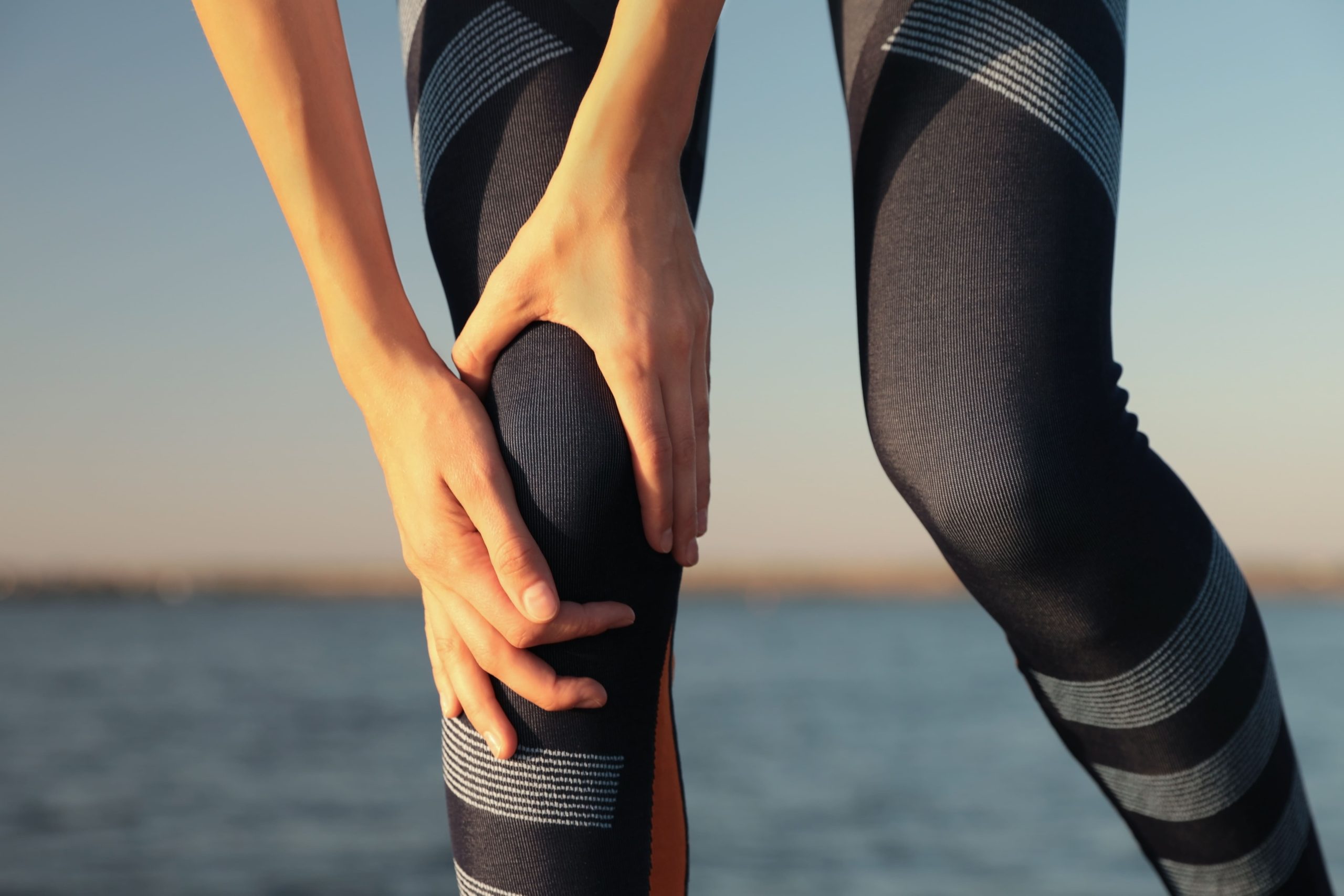
ACL tears frequently happen during non-contact situations. Landing awkwardly from a jump, cutting sharply while running, or suddenly stopping creates forces that exceed the ligament’s tensile strength (its ability to withstand pulling forces). The classic mechanism involves the knee collapsing inward whilst the foot remains planted. This combines valgus stress (inward bending force) with rotation.
PCL injuries require substantial force directed at the proximal tibia (upper shin bone). Motor vehicle accidents in which the knee strikes the dashboard are the most common cause. Sports-related PCL tears occur from falling directly onto a flexed knee or from hyperextension injuries (when the knee bends too far backwards). Isolated PCL tears are less common than combined ligament injuries.
Recognising the Symptoms
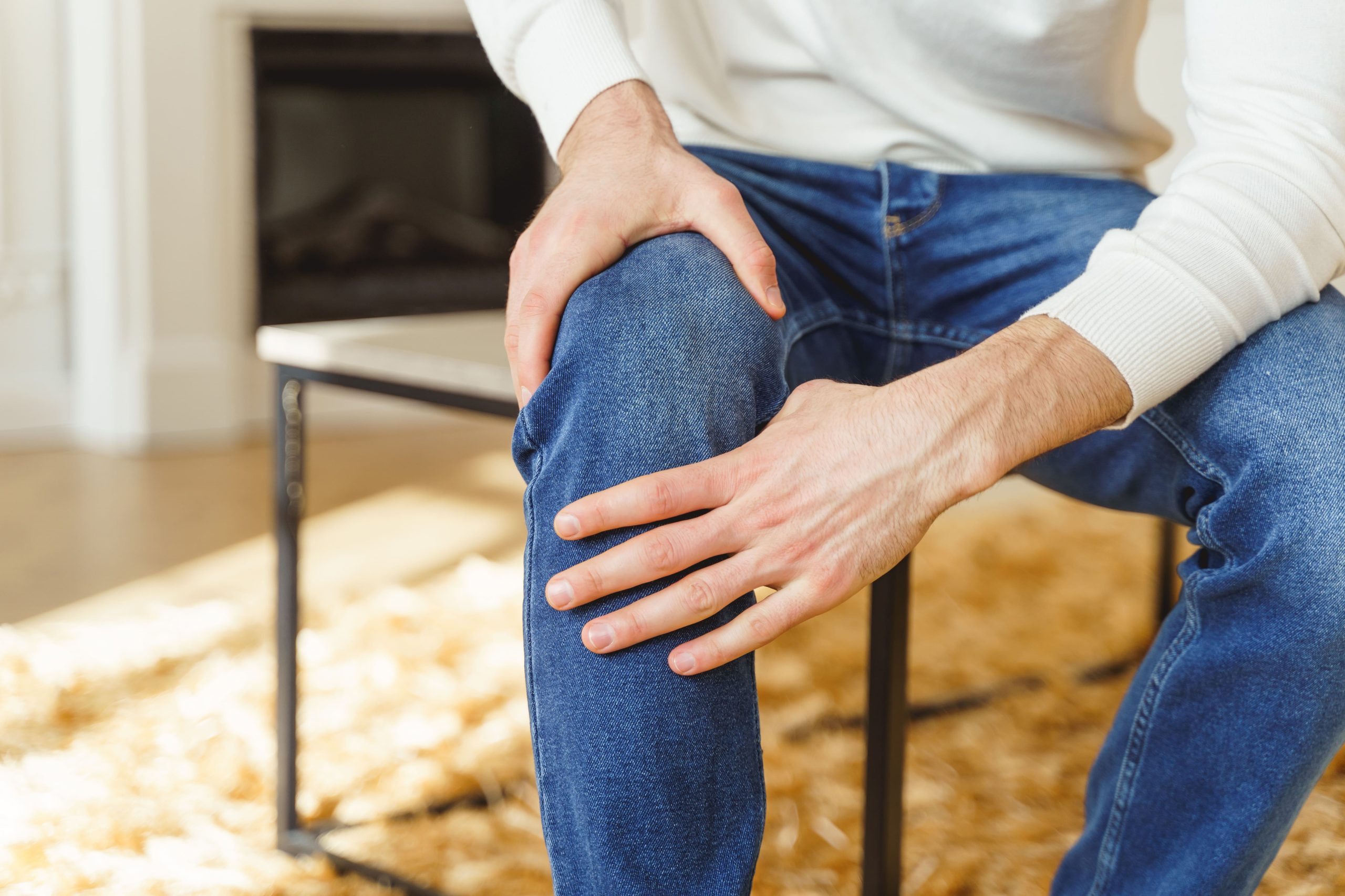
ACL tears often produce an audible pop at the time of injury. Rapid swelling follows within hours. The knee feels unstable, particularly during twisting or pivoting movements. Walking on flat surfaces may feel manageable. Descending stairs or uneven terrain reveals the instability.
PCL tears present more subtly. Swelling develops gradually over days rather than hours. Pain localises to the back of the knee. It worsens when kneeling or walking downhill. The knee may feel “off” without the dramatic giving-way episodes characteristic of ACL tears.
💡 Did You Know?
The ACL hasa minimal blood supply. This explains its poor natural healing capacity. The PCL receives better vascularisation (blood flow) from the synovial membrane (the lining of the joint). Partial tears can sometimes heal without surgery.
Diagnostic Evaluation
The physical examination begins by comparing both knees. The Lachman test (a physical examination technique) assesses ACL integrity by applying an anterior tibial load with the knee slightly bent. Increased translation may indicate ACL disruption. The anterior drawer test evaluates similar movement of the knee in considerable flexion.
For the PCL assessment, the posterior drawer test assesses posterior tibial displacement (movement). The examiner also observes the tibia’s resting position. A posterior sag (backward dropping) compared to the uninjured side suggests PCL insufficiency. The dial test evaluates rotational instability when combined injuries are suspected.
Imaging Studies
MRI (a diagnostic imaging test that uses magnets to create detailed pictures of the inside of your knee) provides a definitive diagnosis. It visualises ligament fibres directly and identifies associated injuries to menisci (cartilage cushions in the knee), cartilage, and other ligaments. Bone bruise patterns on MRI often confirm the injury mechanism. ACL tears typically show bone bruises on the lateral femoral condyle (outer part of the thigh bone) and posterolateral tibial plateau (back-outer part of the shin bone).
X-rays (diagnostic imaging tests that use radiation to create pictures of bones) appear normal with isolated ligament injuries. They remain essential for ruling out fractures. Stress X-rays can quantify the degree of instability. They are taken whilst applying force to the knee. They help monitor healing in PCL injuries managed non-operatively.
Non-Surgical Treatment Approaches
Not every cruciate ligament injury requires surgery. Treatment decisions depend on several factors:
- Injury severity
- Patient activity level
- Associated injuries
- Individual goals
A healthcare professional can provide personalised advice on treatment options based on your specific risk factors. These include your age, activity level, existing medical conditions, and overall knee health.
Initial management for both injuries follows similar principles. Controlling swelling through ice, compression, and elevation takes priority during the first week. Protected weight-bearing with crutches allows pain to settle. Early physiotherapy focuses on restoring the range of motion (how far you can bend and straighten your knee), particularly full extension (straightening). This becomes difficult to regain if neglected initially.
When Non-Surgical Management Works
Isolated PCL tears, especially partial tears, often heal adequately without surgery. The ligament’s blood supply allows scar tissue formation. This provides functional stability for many activities, whilst not restoring normal anatomy. Rehabilitation focuses on strengthening the quadriceps (thigh muscles). These dynamically resist posterior tibial translation (backwards sliding of the shin bone).
ACL-deficient knees can function for daily activities and even some sports in selected individuals. These “copers” develop neuromuscular adaptations (changes in how the muscles and nerves work together). These compensate for ligamentous instability. However, returning to pivoting sports without reconstruction carries risks. Repeated instability episodes can cause meniscal and cartilage damage.
Surgical Treatment Options
ACL reconstruction is a surgical procedure. The surgeon replaces the torn ligament with a graft (a piece of tissue used as a replacement). The graft is typically harvested from the patient’s own tissue (autograft) or using an allograft (donor graft). Common autograft graft choices include:
- Patellar tendon (the tissue connecting the kneecap to the shin bone)
- Hamstring tendons (tissues at the back of the thigh)
- Quadriceps tendon (tissue connecting the thigh muscle to the kneecap)
Each graft type has advantages and considerations. A healthcare professional will discuss these based on your specific situation.
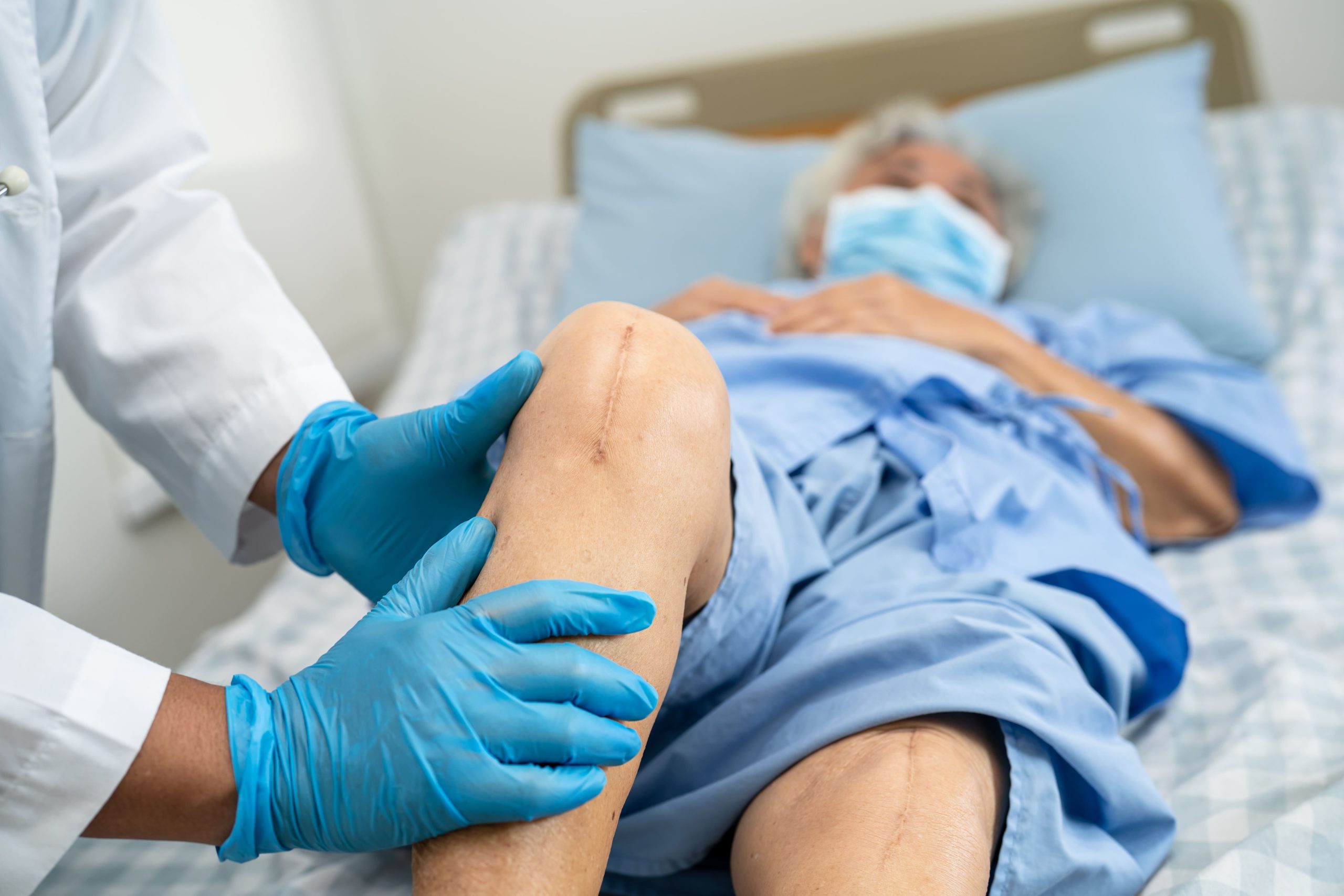
The procedure uses arthroscopic techniques (a minimally invasive surgical approach using a small camera and instruments inserted through tiny incisions). Small incisions provide access for the camera and instruments. The surgeon drills tunnels through the tibia and femur. The graft is positioned to replicate the ACL’s original anatomy. It is secured with various fixation devices (screws or buttons that hold the graft in place). The graft gradually incorporates into the bone tunnels. It undergoes biological remodelling (natural tissue healing and strengthening) over months.
PCL Reconstruction Considerations
PCL reconstruction remains more technically demanding and less predictable than ACL surgery. The posterior approach to the tibia poses risks to neurovascular structures (nerves and blood vessels). Graft tensioning (adjusting the tightness of the replacement tissue) proves challenging. The PCL experiences different forces throughout the knee’s range of motion.
Surgery is generally reserved for specific situations:
- Combined ligament injuries
- High-grade PCL tears with significant instability
- Symptomatic chronic PCL deficiency that hasn’t responded to rehabilitation
Many PCL injuries achieve satisfactory outcomes with structured physiotherapy alone. This applies even to complete tears.
⚠️ Important Note
Combined ACL-PCL injuries (knee dislocations) require urgent evaluation for vascular injury (damage to blood vessels). The popliteal artery (a major blood vessel) runs behind the knee. It can be damaged during severe trauma. Any signs of compromised circulation warrant immediate medical attention.
What Our Orthopaedic Surgeon Says
The decision between surgical and non-surgical treatment involves more than just the MRI findings. We assess how the injury affects your specific activities and goals. Age, occupation, and associated injuries all influence the treatment plan. The goal is to restore function to your life, not simply fix an image on a scan.
Rehabilitation and Recovery Timeline
Post-operative rehabilitation follows a phased approach spanning several months. The biological healing of the graft dictates progression more than pain or functional ability. Rushing rehabilitation risks graft failure before adequate incorporation into bone.
Phase-Based Recovery
The initial phase focuses on protecting the graft whilst minimising complications. Achieving full knee extension, controlling swelling, and restoring quadriceps activation (the ability to contract your thigh muscle) take priority. Weight-bearing progresses gradually, initially with brace support.
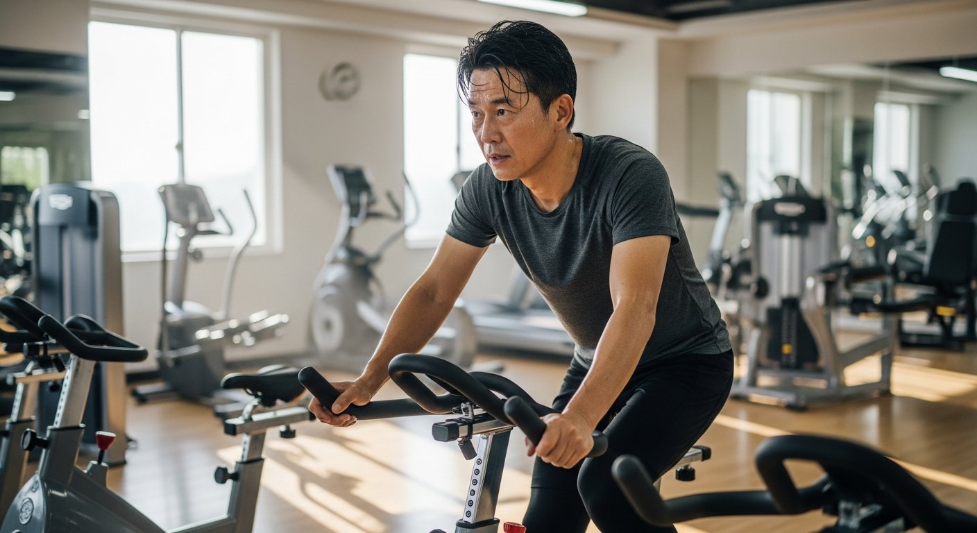
Intermediate phases introduce progressive strengthening, balance training, and controlled range-of-motion exercises. Closed-chain exercises (exercises in which the foot remains in contact with a surface, such as squats or leg presses) stress the graft less than open-chain movements (exercises in which the foot moves freely, such as leg extensions). Cycling, swimming, and straight-line jogging typically begin around three to four months post-surgery.
Return to sport requires passing functional tests. These assess strength, power, and movement quality. Single-leg hop tests compare the surgical and non-surgical legs. They help determine readiness.
Factors Affecting Recovery Outcomes
Graft choice influences early rehabilitation. It shows minimal difference in long-term outcomes. Patellar tendon grafts may initially cause more anterior knee pain (pain at the front of the knee). They provide rigid bone-to-bone healing. Hamstring grafts preserve the extensor mechanism (the structures that allow you to straighten your knee). They require longer incorporation times and may result in some hamstring weakness. Allograft options may be helpful in some individuals with minimal donor morbidity.
Associated injuries significantly impact recovery. Meniscal repairs (procedures to fix torn cartilage cushions) require prolonged restricted motion to allow healing. This extends rehabilitation timelines. Cartilage damage may limit ultimate function regardless of successful ligament reconstruction. Addressing all pathology during surgery, when appropriate, optimises outcomes.
Patient factors influence results. These include compliance with rehabilitation, pre-injury fitness level, and psychological readiness. Fear of re-injury can persist long after the knee has physically healed. This affects return-to-sport rates, independent of surgical success.
Long-Term Considerations
ACL reconstruction reduces but doesn’t eliminate the risk of subsequent knee problems. The contralateral knee (the opposite knee) faces a risk of injury after returning to pivoting sports.
Post-traumatic arthritis (joint degeneration that develops after an injury) can develop in the knees following ACL injury. This occurs regardless of surgical treatment. The initial injury causes cartilage damage that progresses over decades. Maintaining a healthy weight, quadriceps strength, and appropriate activity modification may help slow this progression.
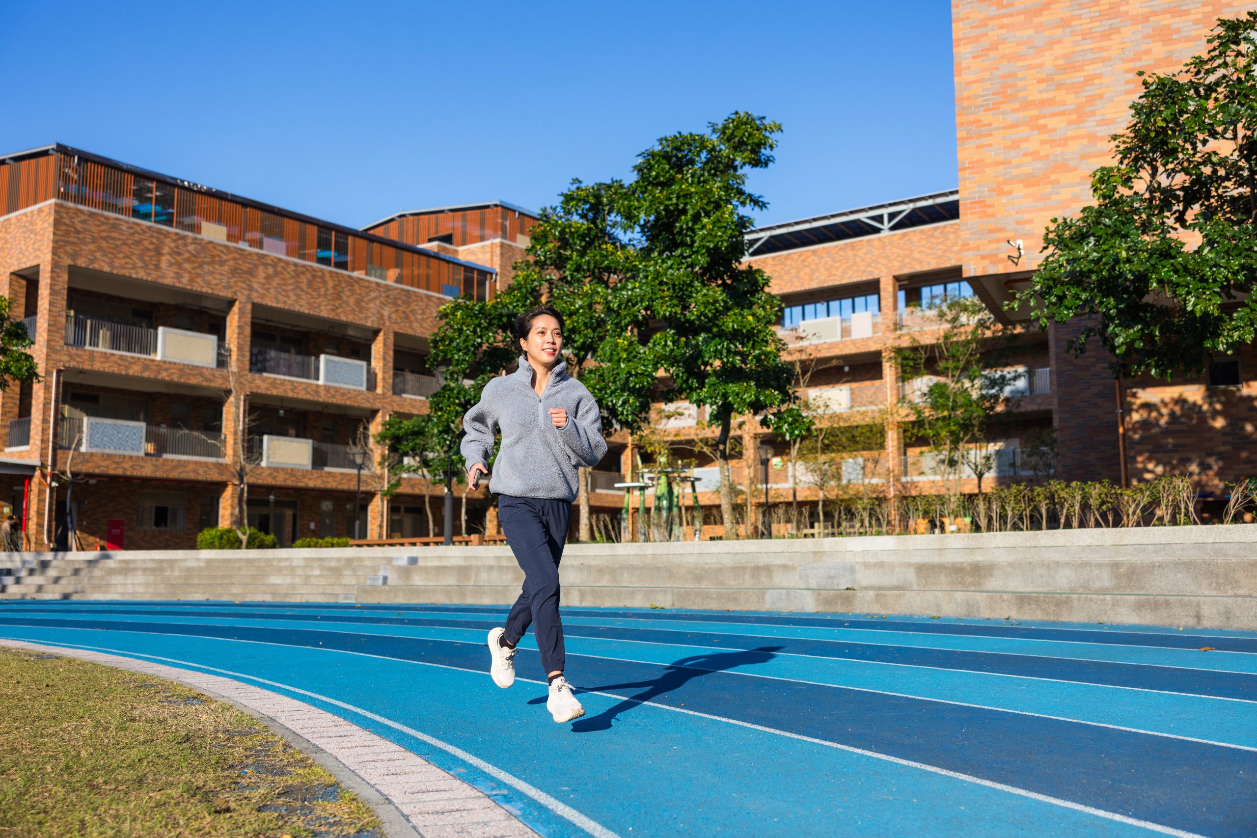
Injury Prevention Strategies
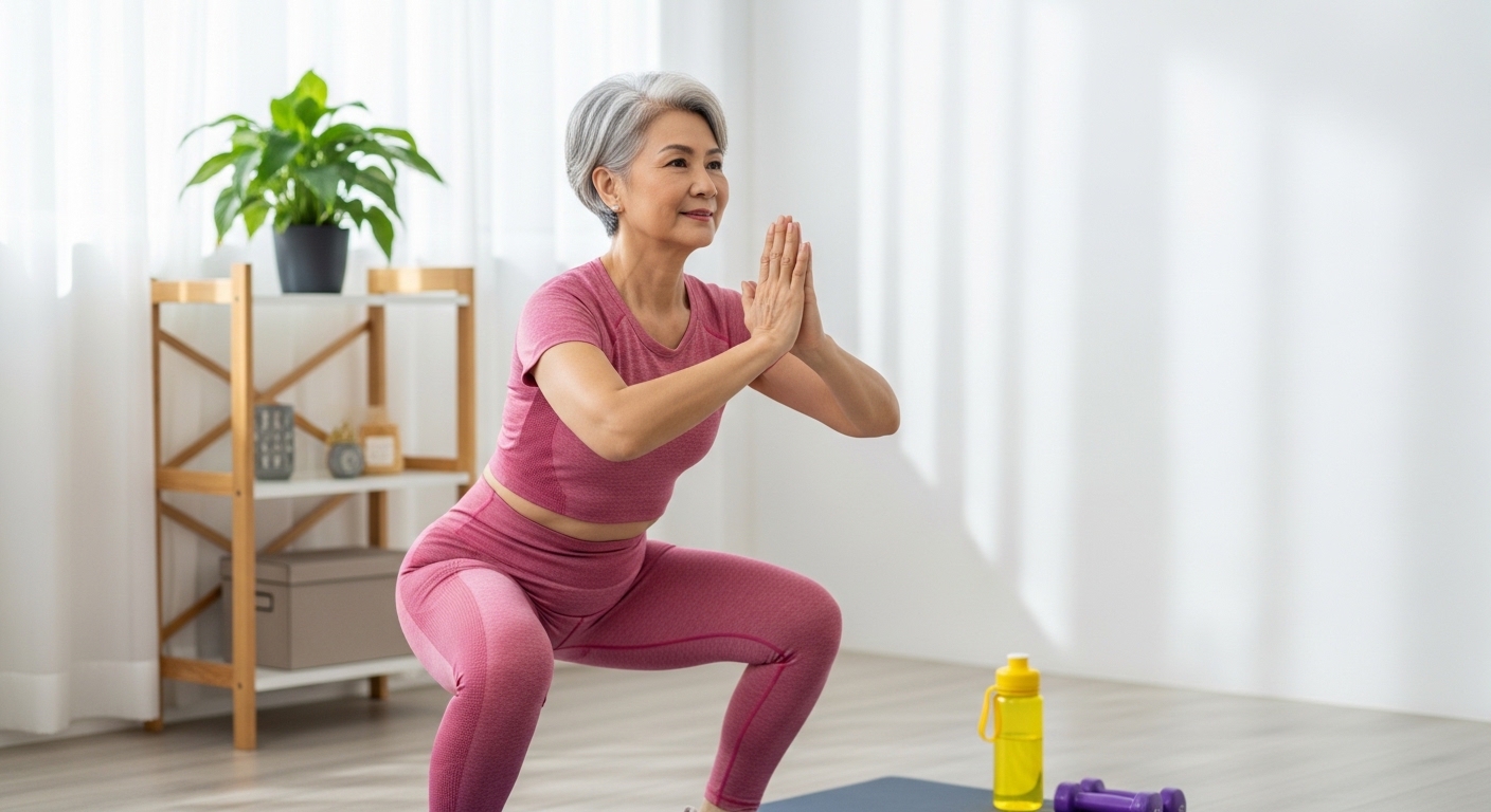
Neuromuscular training programmes (structured exercise programmes that improve how muscles and nerves work together to control movement) can reduce ACL injury rates. These programmes incorporate several elements:
- Balance exercises
- Plyometric training (jumping and landing exercises)
- Movement pattern correction
Consistent participation throughout the season provides greater protection than pre-season training alone.
Movement quality matters more than muscle strength alone. Learning to land from jumps with soft knees, avoiding knee valgus collapse (inward bending), and developing hip and core stability all contribute to injury prevention. Sport-specific training that addresses the demands of your particular activities optimises protection.
✅ Quick Tip
When returning to activity after cruciate ligament injury, start with predictable movements before progressing to reactive situations. Your knee needs to relearn automatic protective responses. These take longer to develop than strength alone.
When to Seek Professional Help
- Immediate severe swelling after a twisting injury to the knee
- Audible pop at the time of injury, followed by difficulty bearing weight
- Sensation of the knee giving way during daily activities or sports
- Persistent instability despite several months of rehabilitation
- Worsening symptoms or new mechanical symptoms, such as locking or catching
- Inability to fully straighten or bend the knee after injury
Commonly Asked Questions
How long after ACL reconstruction can I drive?
Driving typically resumes between four and six weeks post-surgery, though the timeline and degree of recovery vary from person to person. This depends on which leg was operated and whether your vehicle is automatic or manual. Right knee surgery affects brake response time. A healthcare professional will assess when your reaction time and control have adequately recovered.
Will my knee ever feel completely normal after ACL reconstruction?
Individual recovery experiences will differ due to personal health factors. Subtle differences compared to the uninjured knee commonly persist. Some notice mild swelling with heavy activity, occasional knee awareness, or minor strength differences. These typically don’t limit function for daily activities or recreational sports.
Can I avoid surgery if I stop playing sports?
Many people function well with ACL deficiency for daily activities and low-demand recreation. However, instability episodes during unexpected movements can damage menisci and cartilage. The decision involves weighing surgical risks against the cumulative risk of instability-related damage over time. Consult a healthcare professional for advice tailored to your specific situation.
Why does my knee still hurt months after a PCL injury?
Chronic PCL deficiency alters knee mechanics (how the knee moves and functions). This increases stress on other structures. The medial compartment (inner part of the knee) and patellofemoral joint (where the kneecap meets the thigh bone) commonly become symptomatic. Quadriceps weakness allows increased posterior tibial sag (backward dropping of the shin bone). This perpetuates abnormal mechanics. Targeted rehabilitation addressing these factors often improves symptoms. Your recovery timeline may vary based on individual circumstances.
Is ACL tear treatment in Singapore the same as overseas?
Treatment principles remain consistent internationally. They follow evidence-based guidelines. Orthopaedic surgeons in Singapore (doctors who specialise in diagnosing and treating bone, joint, and ligament conditions) train extensively. They maintain familiarity with current techniques and research. Local factors, including rehabilitation access and climate considerations, may influence specific recommendations.
Important Disclaimer: Individual recovery experiences will differ based on personal health factors, injury severity, treatment approach, and adherence to rehabilitation protocols. The information provided here is for general educational purposes and should not replace personalised medical advice. Always consult qualified healthcare professionals for guidance tailored to your specific circumstances and medical history.
Next Steps
Accurate diagnosis determines whether structured rehabilitation or surgical reconstruction is most appropriate. Successful outcomes depend on consistent physiotherapy and gradual progression back to activities. Addressing both the structural injury and neuromuscular deficits optimises recovery.
If you’re experiencing knee instability, giving way episodes, or persistent pain following a knee injury, consult a qualified orthopaedic surgeon to discuss your specific condition and treatment options.