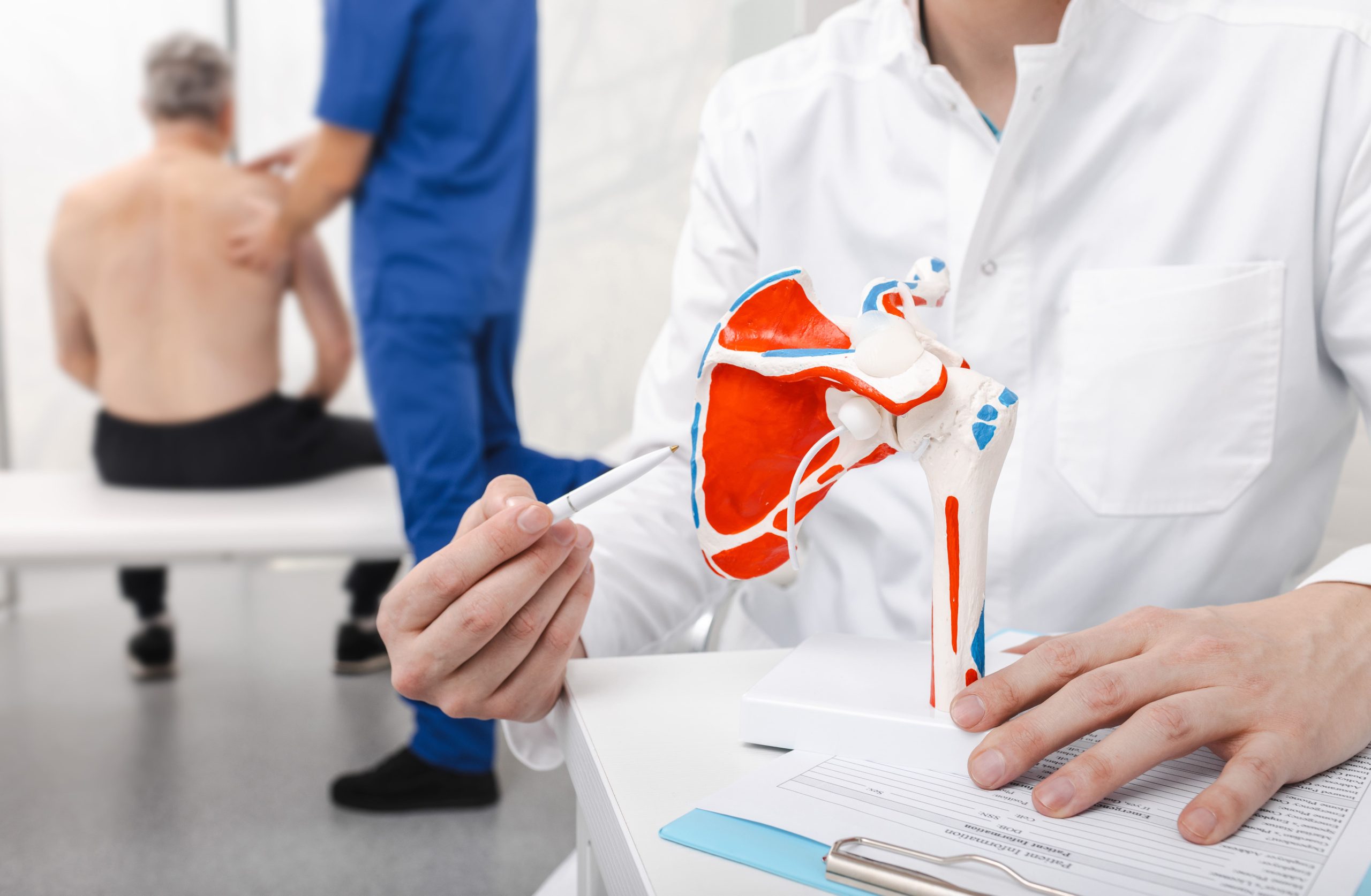
Calcific tendonitis occurs when a specific type of calcium crystal — called carbonate apatite, similar to the mineral found in bone — builds up within the rotator cuff tendons, most commonly affecting the supraspinatus at the top of the shoulder.
The most intense pain tends to occur during the resorptive phase when the body attempts to break down and absorb the calcium. However, many individuals pass through this phase with only mild or no symptoms. Severe acute presentations, while dramatic when they occur, are not universal.
How Calcium Deposits Form in Shoulder Tendons
The exact mechanism triggering calcium deposition remains incompletely understood. However, several factors contribute to its development. The structural cells of the tendon, called fibroblasts, begin behaving like cartilage cells. This causes the tough collagen fibres that make up the tendon to slowly turn into a cartilage-like material — and cartilage-like tissue is capable of forming calcium deposits, which is how the build-up begins.
Three distinct phases characterise the condition’s progression:
Pre-calcific phase: Tendon tissue undergoes cellular changes that create an environment conducive to calcium deposition. Patients typically experience no symptoms during this stage.
Calcific phase: Calcium crystals actively deposit within the tendon. This phase subdivides into formative, resting, and resorptive stages. The formative stage involves ongoing calcium accumulation. The resting stage represents a stable period. The resorptive stage triggers intense inflammation as the body attempts to dissolve the deposits.
Post-calcific phase: Following calcium resorption, the tendon undergoes remodelling and healing. Granulation tissue (new tissue rich in blood vessels) replaces the calcium. It eventually matures into organised tendon tissue.
Alterations in the blood supply within the tendon may contribute to deposit formation. Areas of relative hypoxia (reduced oxygen delivery) appear more susceptible to calcification. The supraspinatus tendon contains a “critical zone” with naturally reduced blood flow. This potentially explains why this tendon develops calcific tendonitis more frequently than others.
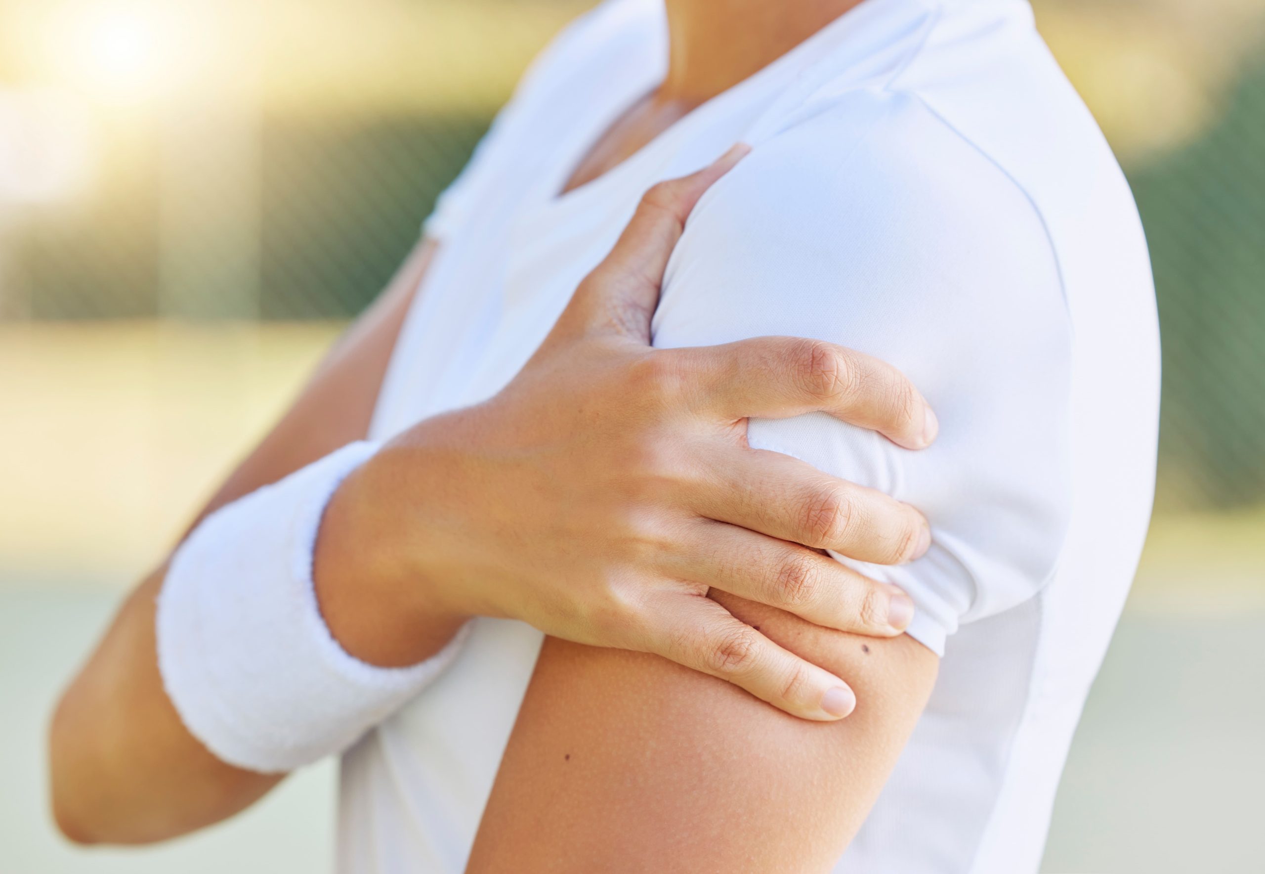
Recognising Calcific Tendonitis Symptoms
Pain patterns in calcific tendonitis vary considerably depending on which phase the condition has reached. During the formative phase, discomfort tends to be mild and intermittent. It is often dismissed as general shoulder strain.
The resorptive phase produces the characteristic acute presentation. Pain can develop rapidly, and many patients report being unable to move the affected arm. They may describe pain that radiates down the arm toward the elbow. Night pain is particularly prominent. Many are unable to sleep on the affected side.
Physical examination findings include:
- Tenderness over the affected tendon, typically at the anterior or lateral shoulder
- Painful arc of movement in a certain range of abduction (when raising your arm out to the side)
- Reduced active range of motion due to pain inhibition
- Weakness in rotator cuff testing, though this often reflects pain rather than true muscle weakness
Chronic calcific tendonitis presents differently. It causes persistent low-grade shoulder discomfort that worsens with overhead activities. Some patients experience recurrent acute flares separated by relatively pain-free intervals lasting weeks to months.
Diagnostic Approaches for Calcific Tendonitis
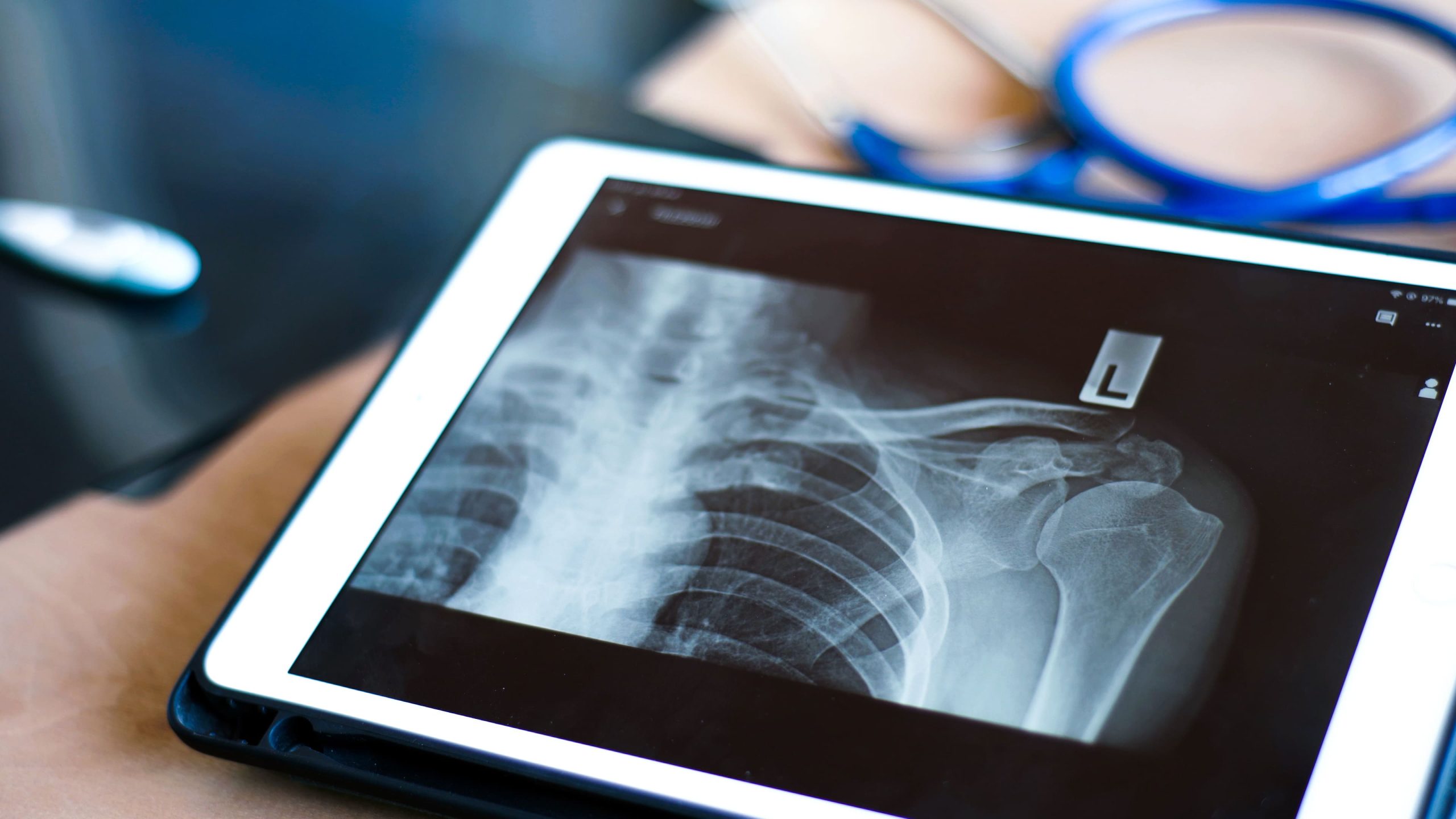
Accurate diagnosis of calcific tendonitis involves several imaging modalities to determine the size, location, and specific stage of the calcium deposits.
- X-rays: Dense, sharply defined spots on an X-ray suggest the deposit is in the formative or resting phase, meaning it is still building up or stable. A cloudier, ‘fluffy’ or poorly defined appearance suggests the resorptive phase, when the body is actively trying to dissolve the deposit and pain tends to be at its worst.
- Ultrasound: This allows for real-time visualisation of the tendon, detects smaller deposits missed by X-rays, and provides guidance for minimally invasive treatments.
- MRI Scanning: While generally not required for a primary diagnosis, MRI is used to evaluate associated issues like rotator cuff tears, bursal inflammation, or bone marrow swelling.
- Clinical Assessment: The number and size of deposits are carefully evaluated, as larger or bilateral deposits may require more comprehensive management or surgical consideration.
Why Some People Develop Calcific Tendonitis
Several factors may increase susceptibility to calcium deposition:
- Metabolic influences: Thyroid disorders, diabetes, and conditions affecting calcium metabolism may increase risk.
- Hormonal factors: Hormonal shifts can influence tendon health and calcium regulation.
- Genetic predisposition: Family clustering suggests that inherited susceptibility and genetic variations in collagen structure may play a role.
- Mechanical factors: While research has not confirmed that repetitive overhead work (like painting or electrical work) causes calcific tendonitis specifically, poor posture and muscle imbalances can create microtrauma that aggravates existing deposits.
Treatment Options for Calcific Tendonitis
Effective management of calcific tendonitis involves a tailored approach that transitions from non-invasive therapies to clinical procedures based on the severity of the patient’s symptoms and the stage of the calcium deposit.
Conservative Management
Initial treatment focuses on reducing inflammation through rest, activity modification, and the use of anti-inflammatory medications. Specialised physiotherapy is then employed to correct muscle imbalances and strengthen the rotator cuff to prevent future irritation.
Corticosteroid Injections
These injections reduce the inflammation and pain around the deposit, which can bring significant short-term relief. However, it’s worth knowing that during the resorptive phase, when the body is actively trying to dissolve the calcium, steroids can actually slow that natural removal process down. Your doctor will weigh up the benefit of pain relief against this trade-off.
Ultrasound-Guided Barbotage
Using real-time ultrasound as a guide, a needle is inserted directly into the calcium deposit. Saline (a sterile saltwater solution) is used to flush out and break up the material — but this works best when the deposit has softened to a paste-like consistency, which typically happens during the resorptive phase. Hard, chalk-like deposits are more resistant to this technique.
Extracorporeal Shockwave Therapy
Shockwave therapy works best when the deposits are still firm and stable — typically during the formative or resting phases. It is generally avoided during the acute resorptive phase, when the shoulder is already severely inflamed, as the treatment can worsen irritation at that stage. Your doctor will assess your X-ray and symptom pattern before recommending it.
Surgical Treatment
When non-surgical methods do not provide adequate relief, arthroscopic surgery may allow a surgeon to locate and remove the calcium deposits through small keyhole incisions. This procedure also provides an opportunity to inspect and repair any concurrent damage to the rotator cuff tendons.
Clinical Perspective on Treatment Timing
Calcific tendonitis frequently resolves spontaneously. However, this provides little comfort during acute episodes. The challenge lies in matching treatment intensity to each patient’s specific situation. Aggressive intervention isn’t always necessary. Neither should patients suffer unnecessarily while waiting for natural resolution.
Timing treatment to the deposit’s phase may improve outcomes. Resorptive phase deposits may respond to image-guided intervention. Stable formative phase deposits may warrant a more conservative initial approach with close monitoring.
Practical Steps for Managing Shoulder Discomfort
Modify aggravating activities: Identify and temporarily avoid movements that reproduce pain. These particularly include reaching overhead or behind the back.
Apply appropriate thermal therapy: During a painful flare-up, ice packs can help calm the inflammation — apply for 15–20 minutes at a time with a cloth barrier. In the chronic, lower-grade phase, gentle warmth can help ease surrounding muscle tension. Avoid applying heat during an active acute flare, as this can increase inflammation and worsen pain.
Maintain gentle movement: Complete rest leads to shoulder stiffness over time, so keeping the joint gently moving matters. Pendulum exercises — letting the arm hang and swing in small, relaxed circles — can help preserve movement without putting stress on the inflamed tendon. During a severe acute flare, check with your physiotherapist or doctor before starting these, as timing matters.
Optimise sleeping position: Avoid lying on the affected shoulder. Place a pillow under the affected arm when lying on the opposite side.
Follow prescribed exercise programs: Physiotherapy exercises address underlying factors. They reduce recurrence risk. Consistency matters more than intensity.
When to Seek Professional Help
- Shoulder pain develops suddenly and severely over hours
- Inability to lift the arm away from the body
- Night pain preventing sleep for more than several days
- Shoulder discomfort is not improving after two weeks of rest
- Pain accompanied by fever or significant swelling
- Previous calcific tendonitis with recurrent symptoms
- Shoulder symptoms following injury or trauma
Commonly Asked Questions
Can calcific tendonitis heal completely without treatment?
Many cases resolve spontaneously as the body naturally resorbs calcium deposits. This process can take months to years, though the timeline and degree of improvement vary from person to person. Predicting which deposits will resolve versus persist remains difficult. Treatment may accelerate recovery and reduce discomfort during the often-prolonged natural course.
Will the calcium deposits return after treatment?
Recurrence after successful treatment does happen, though how often depends on the type of treatment received and individual factors. Addressing contributing factors, such as posture, muscle balance, and repetitive strain, may help reduce the risk of it coming back.
Is calcific tendonitis related to dietary calcium intake?
The calcium in tendon deposits doesn’t come directly from dietary calcium. Reducing calcium intake won’t prevent or treat the condition. Maintaining normal dietary calcium remains important for bone health. The calcification results from local tissue changes rather than systemic calcium excess.
How long does recovery take after barbotage or shockwave therapy?
Response times vary depending on your specific condition. Some patients notice improvement within weeks following barbotage. However, complete resolution may take a few months. Shockwave therapy typically requires longer. Improvement often develops gradually over several months as deposits fragment and resorb.
Can I continue exercising with calcific tendonitis?
Exercise modification rather than complete cessation is typically recommended. Activities that don’t aggravate symptoms can continue. Overhead movements and heavy lifting may require temporary avoidance. Swimming breaststroke, cycling, and lower-body exercises often remain comfortable.
Next Steps
Treatment for calcific tendonitis should be matched to the condition’s phase and severity. Acute resorptive phase presentations—characterised by sudden, intense pain and inability to move the arm—may benefit from prompt intervention. Stable deposits identified incidentally or causing only mild discomfort may warrant initial conservative management with physiotherapy. Accurate diagnosis through X-ray or ultrasound imaging is necessary to determine the deposit stage and guide treatment selection.
If you are experiencing sudden, intense shoulder pain, night pain preventing sleep, or persistent shoulder discomfort not improving with rest, consult an orthopaedic surgeon for clinical examination and imaging to determine whether calcific tendonitis requires treatment.